


")

")
")
")
")
")


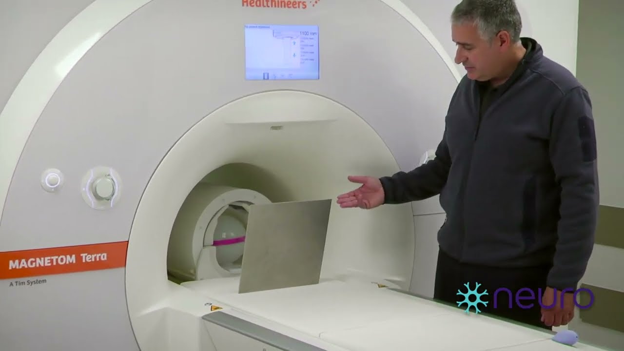
Utilized all throughout the modern healthcare system, magnetic resonance imaging (MRI) is a very strong diagnostic tool. It utilizes an intense magnetic field paired with radio waves to create detailed pictures of the body’s organs and tissues. However, when it comes to going through an MRI scan, having metal in a patient’s body can be dangerous or present difficulties. Different metals like iron, nickel, and cobalt are used in many implants and medical devices; these metals may react unpredictably in the presence of the magnetic field, which could harm the person or change how the image looks. Therefore, before undergoing this procedure, patients must inform their healthcare provider about any metal implants they have had placed as well as shrapnel, bullets, or other pieces of metal that might be inside them. Following correct screening procedures along with advanced imaging techniques will allow professionals within health care settings to ensure safe use while minimizing risk associated with the inherent power found in Magnetic Resonance Imaging machines (MRI).
Understanding Metallic Implants in Relation to MRI Safety
Weighing the risks: metallic implants and the magnetic field of an MRI
In terms of strength, no other operative procedure comes close to that of a magnetic resonance imaging (MRI) scan. As such, metal implants could significantly jeopardize the safety and efficiency of this process through their interaction with its powerful magnetic fields. Ferromagnetic substances such as iron, nickel, and cobalt, which make most implants, are easily influenced by these forces because they have high permeability rates for magnetism. The outcome may be heating within or around the implant, leading to tissue injury at that site; also, magnetism can cause immediate physical harm by moving or displacing ferromagnetic objects against the patient’s body. Even metals not classified as ferromagnetic can still distort images obtained during an MRI by producing artifacts that hide details about scanned areas, thus affecting correct diagnosis too much reliance on the accuracy. Therefore, it is important for doctors to look into patients’ histories so as to detect the presence of any metallic material before scanning them, lest we put their lives in danger while trying to save our own lives using diagnostic tools like MRI scans.
The details of ferromagnetic substances and their impact on MRI scans
Unique interaction with MRI scanners is caused by the powerful magnetism of ferromagnetic materials like iron, nickel, and cobalt. This can lead to several problems when these substances are exposed to a magnetic field generated by an MRI system:
- Heating: The particles within ferromagnets vibrate rapidly as they align themselves with or against the magnetic field lines – creating heat in the process. If this occurs near sensitive tissues or other heat-sensitive organs, it could cause severe burns or even damage them.
- Movement: It is important that any object made out of a ferromagnet be securely fastened so that it does not move during imaging; otherwise, such an item might get attracted towards magnets due to strong attraction forces created by those fields surrounding its environment, thereby leading not only harm but also injury anybody who comes close enough.
- Image Distortion: Ferrous metals can create large artifacts on images taken using an MRI machine which will make diagnosis hard if not impossible sometimes. This happens because scanning machines use powerful magnets to map hydrogen atoms’ location inside our bodies – something interfered with by these metals’ magnetic properties.
Knowing about these dangers is crucial for patients and medical staff alike when dealing with magnetic resonance imaging devices. Such awareness helps reduce risks involved during such examinations, thus making them safer while increasing their effectiveness as diagnostic tools.
Advice and safety measures for patients with metal implants
There are certain precautions that must be taken by the patient who has a metal implant before getting an MRI scan so as not to compromise their safety or image quality. Here are some things they must do:
- Inform the Technician: Prior to starting magnetic resonance imaging (MRI), it is important to tell the healthcare provider about any metal that is within one’s body; this includes shrapnel or other metallic objects. Medical devices such as pacemakers, cochlear implants, clips made from metals and artificial limbs which contain joints should also be mentioned.
- Provide Documentation: Where possible present documents showing what type of metals were used during surgery so that doctors can determine if there would still be need for further evaluation on how compatible such materials are with MRI machines’ powerful magnetic field. Majority of current day implants have been designed in such way that they will not interfere but it is good practice to verify this.
- Go Through Evaluation: Patients should undergo thorough assessments in order for surgeons planning an operation around them to not only know their location but also understand the different types used. By doing this, chances of causing injury or distorting images produced by scans can be minimized while establishing which areas need closer attention.
- Follow Special Instructions: There might be additional requirements given by healthcare providers like fasting prior to tests or changing medication schedules; however, sometimes another safer method without exposure limitations due to metals could be suggested.
These steps ensure better diagnosis through imaging while protecting personal health, too.
Can You Get an MRI with Dental Fillings, Stents, or Pacemakers?

MRI Compatibility of Dental Fillings
In general, MRI can be done on people who have dental fillings made of amalgam composite or any other material. Metallic as they may be, amalgam fillings do not seem to affect the quality of an MRI image significantly because only small quantities consist of stable substances are used. Also made from plastic compounds and ceramics mostly, composite fillings are safe for use in an MRI environment, too; they don’t compromise safety during this procedure, nor do they affect its outcome. Nevertheless, it would still be wise for patients with such conditions to let their dentists know about them before going through scans so that appropriate advice and care can be given by radiologists.
Evaluating MRI Safety of Common Medical Devices: Stents and Pacemakers
Stents are used to keep arteries from getting narrow or blocked. There are different kinds of stents, some of which can be used with magnetic resonance imaging (MRI). Whether a stent can go in an MRI machine depends on what it is made from and how long it has been inside the body. Most new stents will not be harmed by an MRI scanner if certain conditions are met. These usually involve waiting a specific amount of time after putting in the stent, often about six weeks. Patients should know the exact type and model number of their stent so they can get accurate advice from their doctor.
Pacemakers help control irregular heart rhythms by sending electrical signals to the heart muscle. Because of these signals, people with pacemakers have not generally been allowed near MRI scanners in case the magnets inside the machine interfere with this electronic equipment or cause other health problems. But now, there are special pacemakers that are safe to use during an MRI scan. They do not break when they enter strong magnetic fields as long as certain conditions, such as specific field strengths, are met.
For both types of medical devices — stents and pacemakers — you must look at four things: which device exactly is being used (model number), what material is it made out of (composition), how long ago was it implanted (time since implantation), and where will this test take place (specifically magnetic strength environment). Patients need to inform their healthcare provider and MRI technologist about any implants before scheduling an exam to ensure patient safety throughout the procedure.
Enhancements in safe-for-MRI pacemakers and stents
Safe-for-MRI pacemakers and stents are among the most significant developments made by the medical community. The goal was to ensure that patients with these implants could safely undergo an MRI scan. Stents that are safe for use during MRI scans have been made from non-ferromagnetic materials, so they do not move or heat up when exposed to magnetic fields. Similarly, advancements in pacemaker technology created MRI-conditional devices that work normally within an MRI environment, provided some conditions are met. These innovations include better designs for leads, programming options that minimize interference as well as scanning parameters that guarantee the safety of operation of such devices. Such improvements mark a significant shift in care towards patients; they enable full diagnostic imaging while retaining life-saving device functionality.
Dealing with Near Metal Implants During MRI Scans

Approaches to Decrease Picture Distortion Around Metallic Implants
To accurately diagnose patients, it is crucial to minimize image distortion near metal implants during an MRI scan. Here are some things that radiologists and MRI techs do so they can see better:
- High-Bandwidth Pulse Sequencing: This method reduces the spatial extent of susceptibility artifacts around metal implants, which distort images taken during the scans.
- Selecting Imaging Planes: The most complicated areas benefit greatly from improving image quality by aligning an imaging plane perpendicular to its longest axis when implant is involved with it.
- Metal Artifact Reduction Sequences (MARS): MARS has been created specifically to address how an MRI looks vis-a-vis metallic implants. They adjust pulse sequence parameters in order to bring about a decrease in artifacts as well as increased visibility around these tissues on scans themselves.
- Slice Encoding for Metal Artefact Correction(SEMAC): SEMAC is a new technique that uses high-bandwidth pulses combined with view-angle tilting to correct both through-plane and in-plane artifacts. It includes extra z-axis encoding steps which compensate for distortions caused by metals thereby providing more accurate depiction of anatomy near metals.
- View-Angle Tilting (VAT): VAT compensates for the in-plane distortion caused by the presence of metal. In this case, magnetic field gradient is tilted such that it aligns itself with distorted magnetic field around an implant hence neutralizing any form of distortion brought about by metals.
Taking into account these strategies will allow medical professionals to improve significantly on the quality of MRI scans done on patients having metallic implants thus leading to better diagnosis and care for them.
Apprehending static and powerful magnetic fields’ part in imaging quality
In MRI technology, one of the most important factors that determine how well an image will turn out is its static magnetic field (B0) strength. This establishes a foundation for the alignment of nuclear spins within our bodies. The signal-to-noise ratio (SNR) increases with stronger fields measured in Tesla units, which directly translates into clearer and more detailed images being produced. Conversely, strong gradient magnetic fields are modulated during scans so they can encode spatial information about where these signals came from inside the body; this enables their accurate localization. Rapidly switching them defines resolution as well as speed of imaging, thus allowing us to see complex structures in detail. Therefore, the interaction between such magnets should be optimized for better qualities of MRI, leading to diagnosis improvements.
Recent improvements in MRI technology to better accommodate metal implants
Recent developments in MRI technology have greatly improved imaging capabilities for people with metal implants. These changes aim to reduce the amount of ‘noise’ created by such objects and detected by the scanner.
1. Methods of High-Resolution Scanning: Ultra-high-resolution scanning techniques improve the clarity of images taken near metal implants. By capturing finer details, these techniques can help minimize blurring caused by metallic objects.
2. Metal Artifact Reduction Sequences (MARS): Software algorithms that process MRI data through a series of Metal Artifact Reduction Sequences have been updated so as to negate much of the distortion caused by metals on resultant pictures. This allows for clearer views of soft tissues around prosthetic devices.
3. Multi-Frequency Imaging (MFI): MFI scans an area at more than one frequency, making it easier to distinguish between artificial and natural materials within said region. Consequently, shadows cast by metallic things are reduced, and a more accurate representation is obtained.
4. Advanced Magnet Designs: Designing stronger magnets or using higher Tesla ones makes images crisper because they can differentiate between various types of body parts even if there are different metals present.
5. Customizable Gradient Fields: Modern gradient coil technology now permits operators to tailor gradients directly over areas where there is likely going to be distortion caused by the implant material; this results in more precise images due to cancellation effect produced after interfering waves cross each other inside vicinity.
These advances are all about accuracy. Accuracy when diagnosing patients who’ve had joint replacements done or any other kind procedure involving the use of steel screws etc., thus avoiding wrong conclusions which might lead either missed treatments or unnecessary surgeries altogether
Identifying and Managing Metal in Your Body Pre-MRI
Pre-examination screening for metal objects and devices in patients
Among the many steps taken before an MRI, one of them is to screen a patient’s body for any metallic objects or devices. This is done because it ensures the safety of the patient and the accuracy of the results obtained from the magnetic resonance imaging. The following are some key processes involved during this screening:
- Questionnaire: Patients are required to fill out a detailed questionnaire where they state if they have ever had surgery before or been implanted with anything among others, such as shrapnel, tattoos, etc., which might contain metals.
- Physical examination: A doctor may carry out specific physical examinations especially when there is indication from what was filled on forms by patients about having had implants made from metals inside their bodies.
- Reviewing medical records: Records of the patients are checked to see if there is any known implant like pacemakers, cochlear implants or even metal plates used during surgeries that might not be remembered by individuals themselves.
- Metal detector screening: At times handheld detectors may be used in some hospitals mainly as additional measures for individuals who have high chances of having small metallic fragments within them due to various reasons.
- Consultation with Radiologist/Technician: Wherever necessary, further discussions between an MRI technician/radiographer shall take place so as to assess the risks involved based on identified potential problems and decide whether modifications should be made towards imaging or another diagnostic approach adopted instead.
If these steps are followed strictly then providers will lower chances of occurrence hazards hence making sure that both person being scanned through MRI machine remains unharmed while accurate results are achieved.
The risks of bringing metal into the MRI room
The MRI machine’s strong magnetic field makes it dangerous to bring any metallic objects into the scanning area. Objects made of metal can be turned into projectiles by this magnetic field thereby endangering the patient as well as technicians and causing damage to the MRI equipment itself. Also, metal within a person’s body, such as implants or fragments, can be displaced or heated up by this magnetic field, thus leading to internal injury. The magnetic force produced by an MRI scanner may interfere with electronic devices like pacemakers implanted in people’s bodies, which may result in serious health consequences if left undetected; therefore, strict screening for metals is necessary for safety purposes during the diagnosis process.
What should I do if am not sure whether there is metal inside me or not?
It is important to let your doctor know about any doubts concerning the presence of metals in your body before going for a Magnetic Resonance Imaging (MRI) scan. X-rays or other imaging tests might be advised first so as to rule out hidden fragments or implants that could complicate matters during an MRI procedure. You need to disclose all information regarding previous surgeries and incidents involving metallic items; this will enable them to take necessary precautions aimed at protecting you while ensuring accurate diagnosis without complications from an examination that uses powerful magnets
The Influence of Metal Implants on MRI Quality

Determining what causes image artifacts in magnetic resonance imaging
MRI scans of patients with metal implants are often characterized by poor quality, which usually results from artifacts hiding the clarity and accuracy of the scans. The reason behind these artifacts is that metals distort the magnetic field needed for generating MR images. This creates signal loss as well as geometric distortion around regions being imaged due to variations in magnetic fields near implanted metals. The severity of this effect may differ depending on factors such as the size or shape of a given implant together with its location inside the human body, among others. Notably, ferrous metals are more vulnerable to strong magnetism, hence causing bigger errors. Radiologists must understand these relationships if they are to correctly interpret MRIs, especially in individuals having artificial joints; sometimes, this calls for special techniques that limit such distortions during scanning procedures.
The effect of metal implants on the quality of magnetic resonance imaging
When there are metal implants inside a person’s body, it can greatly affect the outcome of Magnetic Resonance Imaging (MRI). This influence exhibits itself as artifacts—distortions or breaks in the image—that may make the scan useless for diagnosis. The strength with which this happens depends on certain factors such as what magnetic properties the implant has got, its size, and where it is situated relative to the area under investigation. Though still able to disturb the scanning process, non-ferrous metals generally cause less distortion than ferrous ones, which react strongly with MRI’s magnetic field. Despite various advanced imaging techniques and sequences having been developed so far to deal with these problems, however, metals remain one of the hardest obstacles to achieving clear and accurate diagnostic MRIs.
Methods of overcoming challenges caused by metallic objects
Lately, the quality of scans on patients with metal implants has been greatly improved through MRI advancements. These discoveries are aimed at reducing the artifacts produced by metallic objects, therefore ensuring a clearer and more accurate imaging outcome. They include:
- High-Bandwidth Pulse Sequences: These sequences minimize the interference caused by metal hence reducing artifacts by altering the frequency at which an MRI machine operates.
- View Angle Tilting (VAT): In this case, the angle of an MRI’s magnetic field is adjusted relative to a metal implant so as to mitigate against any image distortions.
- Metal Artifact Reduction Sequences (MARS): MARS refers to specialized imaging techniques that have been designed for detecting and correcting distortion as well as signal loss brought about by metals.
- Slice Encoding for Metal Artifact Correction (SEMAC): SEMAC is among those methods which add extra steps into an imaging process in order to specifically deal with distortions caused by metal implants.
These technological solutions play a critical role in enhancing diagnostic accuracy of MRI scans for individuals who have these devices fixed in their bodies thereby enabling radiologists make more precise judgments when interpreting such findings.
Navigating MRI Procedures with Surgical Clips and Other Metal Devices

Anesthesia stitches and magnetic resonance imaging: patient knowledge requirements
Surgical clips may cause anxiety in individuals who are required to undergo an MRI as a result of reports regarding magnetic interference and the creation of artifacts. The majority of modern-day surgical clips are made from materials that can be used during an MRI, for example, titanium, which reduces the chances of magnetic interference. Before scheduling an MRI scan, it is important for patients to inform their physician or radiologist about any previous surgeries involving metal implants, including those with anesthesia stitches. The healthcare provider will then check if these clips have been manufactured using components that will not react negatively when exposed to magnets such as those found in MRI machines. In cases where there are older models or unknown types of clips, specific measures might be taken, like lowering the strength of magnetism or adopting more advanced methods for imaging so as to mitigate the risks involved, but this happens very rarely does it happen? If necessary precautions are taken into account, then can we say that, most times, does it require further investigation at all? Is there anything else I should add here…?
The safety concerns of MRI scanning for people with implanted metal objects
The main issue in the safety of implanted metal devices before being put under an MRI scan is that they may move or get heated by magnets, causing injury to tissues or interfering with their normal functioning. A comprehensive evaluation should be done on what type, where it was placed, and its material composition, among other things, so as to know how risky this can be. Non-risky items are those which have been labeled “MRI safe” by manufacturers only. For “MRI conditional” items, lower magnetic field strengths may be used during scanning sessions, but such instructions must come from makers, too. If there is no indication of whether an implant is safe or not, then doctors need to make sure that hosting such procedures could still work well without harming patients even further through communication while taking into account every other metallic object within one’s body because patient health should always be protected against any possible harm even as we maximize the usefulness of diagnostic capabilities entailed in Magnetic Resonance Imaging (MRI).
When can I have an MRI if there is metal in my body?
To figure out whether it is safe to undergo an MRI scan with metal in the body, you need to know what type of metal it is and where it’s located, as well as some information about the procedure itself. In general terms, this means that if a device has been implanted into a person’s body and labeled “MRI-safe” or “MRI-conditional” by its maker, then yes —there should be no problem letting such patients undertake magnetic resonance imaging examinations since these items do not create any known risks under specified conditions of use during scanning. Apart from this, when we talk about stuff being non-ferromagnetic (not attracted by magnets) and fixed firmly in tissues like some kinds of surgical clips or dental fillings, there may hardly be any danger at all. A patient should inform doctors about his or her complete implant history, including the presence of shrapnel, among other things, before going for a check-up that requires machines with strong magnetic fields, such as those used in hospitals for carrying out scans on people’s bodies. It is important, therefore, that one works closely with their healthcare provider, who will work together with radiologists and manufacturers of these devices so as to establish necessary safety precautions based on individual cases before allowing them to go through an MRI examination.
Reference sources

- “MRI Safety Guidelines for Patients with Metal Implants” – Radiology Journal
- Source Type: Academic Journal
- Summary: This academic journal outlines comprehensive MRI safety guidelines for individuals with metal implants, addressing the complexities of undergoing magnetic resonance imaging procedures. It classifies different types of metal implants and provides essential recommendations to ensure patient safety during MRI scans.
- “Navigating MRI Procedures with Metal In Your Body” – Healthcare Blog Post
- Source Type: Blog Post
- Summary: This blog post offers practical insights into navigating MRI procedures when metal is present in the body, discussing the challenges and considerations involved. It defines the risks associated with metal objects in MRI environments and provides tips to enhance safety and optimize imaging outcomes.
- MRI System Manufacturer Website – Safety Information Section
- Source Type: Manufacturer Website
- Summary: The safety information section on a reputable MRI system manufacturer’s website offers valuable resources on ensuring MRI safety with metal in the body. It compares different metal types affecting MRI compatibility, provides technical classifications, and offers recommendations for healthcare professionals and patients undergoing MRI scans with metal implants.
Frequently Asked Questions (FAQs)
Q: If I have metal in my body, can I get an MRI?
A: Its safety depends on what kind of metal it is and where it is located. There are metals that can be used in MRI scans safely while some may cause harm. Make sure you tell your doctor about any implant before going through with an MRI.
Q: What should someone with metal inside them do if they need an MRI?
A: Let the healthcare provider know about all implants or objects containing metal within your body. They shall evaluate whether an MRI is safe or not by considering factors such as the type of metallic substance used, its location and purpose behind this examination.
Q: What are the dangers of metal in an MRI room?
A: Anything made of metal can be a perilous object to have in an MRI space because it can act as a projectile when it gets into the magnetic field. Observing safety regulations and clearing off anything metallic before going into the room is very important.
Q: Is it safe to use objects containing metal, such as dental fillings or clips, for aneurysms during an MRI scanning session?
A: While certain metallic things are suitable for being scanned by an MRI machine, there are those which cannot be used due to their non-compatibility with this device. In order to find out if it’s safe or not, consult your doctor as well as the technician who will operate on you.
Q: What if I have metal in my body and need an MRI?
A: Before going into the room where the scan is done, tell your healthcare team about any metal you may have. They will advise you on what to do.
Q: In MRI scans that involve metal implants, what does RF (radio frequency) do?
A: It creates images of the body. But with metals around, there can be interaction between them and radiofrequency which causes artifacts in the images. Healthcare workers take these into mind while doing MRI scans with metallic objects inside the patient’s body.
Q: Can I safely use contrast agents for MRI if I have metal in my body?
A: Yes, they can be used as long as there are no known allergies or contraindications to their use. However, it is still important that patients let their healthcare provider know about any preexisting metals before receiving contrast agents for magnetic resonance imaging procedures.
